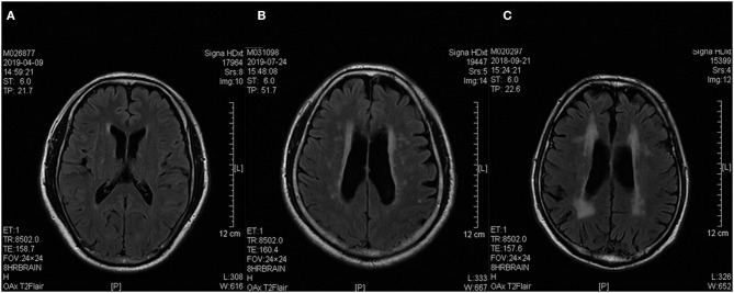
Figure 1.
Sample brain T2 FLAIR MRI images of three patients. (A–C) Based on the Fazekas' score, Patients (A–C) were rated as 1, 2, and 3, respectively. They were grouped into the low burden SVD, moderate burden SVD, and high burden SVD groups, respectively. FLAIR, fluid-attenuated inversion recovery; MRI, magnetic resonance imaging, SVD, small vessel disease.

