Figure 9.
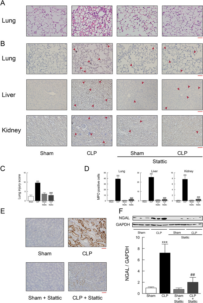
Effect of stattic treatment on tissue injury in mouse tissues following CLP-induced sepsis. Lung, liver, and kidney tissues were harvested 18 h after surgery. (A) Lung tissue sections stained with hematoxylin and eosin. (B) Lung, liver, and kidney tissue sections stained with antibody against MPO followed by peroxidase staining. MPO-positive cells were stained brown and indicated by red arrowheads. Scale bar, 100 μm. (C) Semiquantitative analysis of lung tissues by lung injury, which was assessed by scoring from 0 to 4 as described in Methods (n = 6). (D) Quantitation of MPO-positive cell counts in lung, liver, and kidney tissues. The average of MPO-positive cell number in five fields per sample was calculated (n = 6). (E) Kidney tissue sections stained with anti-NGAL antibody followed by peroxidase staining. Scale bar, 100 μm. Shown are representative images from three independent experiments. (F) Western blots of NGAL protein expression. Typical blots are shown in the top traces. NGAL protein levels were normalized to GAPDH (n = 6). ***P < 0.001 compared with the respective control (18 h after sham operation). ##P < 0.01 and ###P < 0.01 compared with CLP alone.

