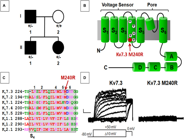
FIGURE 1.
Pedigree, schematic representation of a single Kv7.3 subunit, and functional analysis of mutation at position 240. (A) Pedigree of the family. (B) Topological representation of a single Kv7 subunit. Arrows indicate the location of the mutation investigated (shaded in red). (A–D) Indicate the four putative alpha-helical domains identified in the Kv7 C-terminal region. (C) Sequence alignment of the S4 segments of the indicated Kv subunits (www.ebi.ac.uk/Tools/psa/). Residues are colored according to the following scheme: magenta represents basic; blue represents acid; red represents non-polar; green represents polar. 1, 2, 4, 5, and 6 refer to the positively charged arginines numbered according to their position along the S4 primary sequence. (D) Macroscopic currents from the indicated homomeric channels. Current scale, 50 pA; time scale, 0.2 s.

