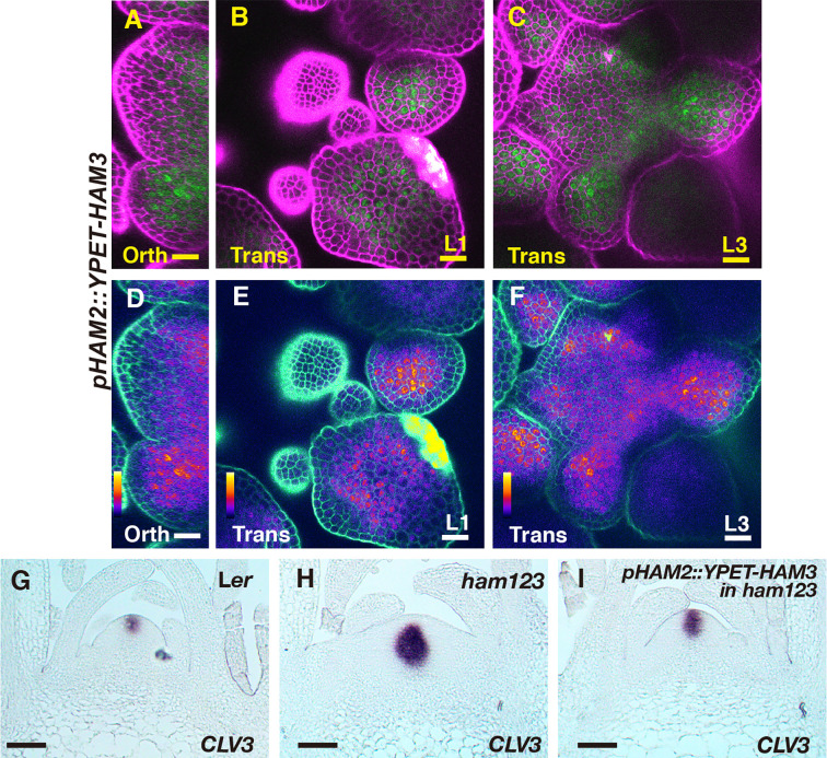
Figure 5.
The expression and function of pHAM2::YPET-HAM3 in the SAM. (A–F) Confocal imaging of a pHAM2::YPET-HAM3 translational reporter in a SAM of ham123 from orthogonal view (A, D), transverse view in L1 (B, E), and corpus (C, F). (A–C): merged channels from YFP (green) and PI (purple). (D–F): merged channels from the quantified YFP (quantitatively indicated by color) and PI. Color bar: Fire quantification. Scale bar (A–F): 20 µm. (G–I) RNA in situ hybridization of CLV3 in the SAMs of wild type (Ler) (G), ham123 (H), and pHAM2::YPET-HAM3 in ham123 (I) at the same developmental stage (27 DAG). Scale bar (G–I): 50 µm. At least three biological replicates were performed for each genotype with similar results.

