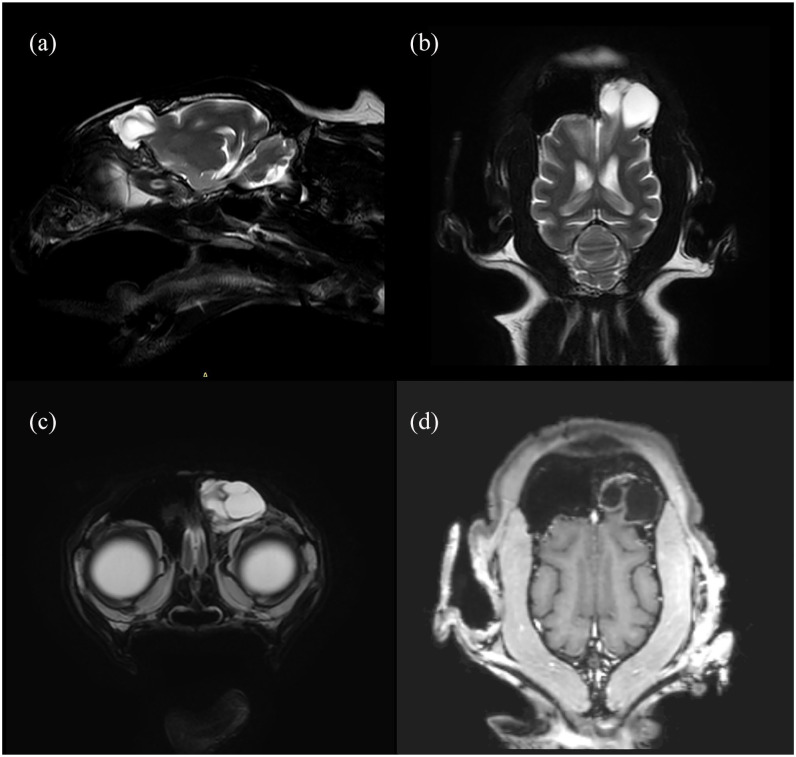
Figure 3.
T2-weighted (a) parasagittal, (b) dorsal and (c) transaxial MRI, and (d) T1-weighted post-gadolinium dorsal MRI of a cat after transfrontal craniotomy. (a) Herniation of a fluid-filled lesion containing small structures of brain tissue into the frontal sinus. (b) The boundaries of the lesion are continuous with the brain parenchyma. A thin septum originating from the cerebral cortex extends throughout the lesion and terminates with a broader-based attachment at the wall of the lesion. (c) The lesion fills the entire left frontal sinus. (d) Mild contrast uptake at the margins of the lesion facing the frontal sinus wall

