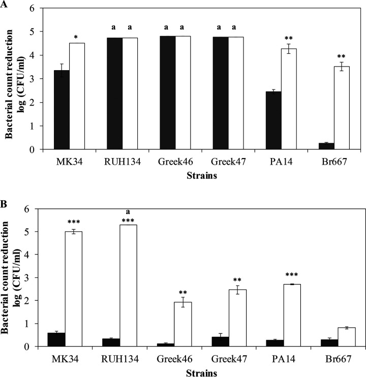
FIG 5.
Antibacterial activity of LysMK34 against different A. baumannii (MK34, RUH134, Greek46, and Greek47) and P. aeruginosa (PA14 and Br667) strains. The bacterial cells were resuspended in a buffer with either low tonicity, corresponding to a high intracellular turgor pressure (20 mM HEPES-NaOH [pH 7.4] alone or supplemented with 0.5 mM EDTA; dark gray and white bars, respectively) (A), or high tonicity and thus a low intracellular turgor pressure (20 mM HEPES-NaOH, 150 mM NaCl [pH 7.4] alone or supplemented with 0.5 mM EDTA; dark gray and white bars, respectively) (B). Antibacterial activity is expressed as the reduction in the bacterial number in log(CFU/ml) after exposure to LysMK34 (25 μg/ml in buffer with low tonicity and 500 μg/ml in buffer with high tonicity) for 2 h. Each value represents the mean ± the SD of three independent replicates. Asterisks represent statistical differences compared to cells not treated with 0.5 mM EDTA (Student t test [*, P < 0.05; **, P < 0.01; ***, P < 0.001]). An “a” label indicates a reduction in the bacterial number that is under the detection limit (10 CFU/ml).

