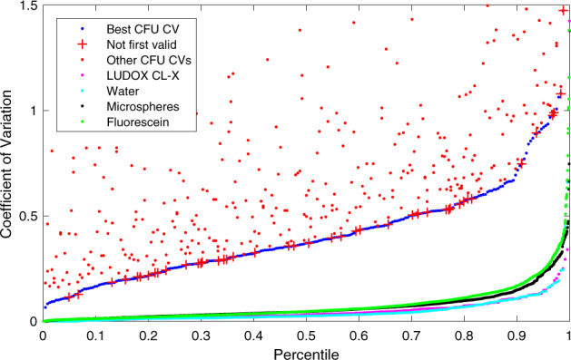
Fig. 2. Distribution of the coefficient of variation for valid replicate sets in CFU, LUDOX/water, microspheres, and fluorescein (all teams included).

CFU models are generated from only the best CV dilution (blue); other dilutions are shown separately above. Even the best CV CFU dilutions, however, have a distribution far worse than the other four methods, and are surprisingly often not the lowest dilution (red crosses). Of the others, LUDOX (magenta) and water (light blue) have the best and near-identical distributions, while microspheres (black) and fluorescein (green) are only slightly higher.
