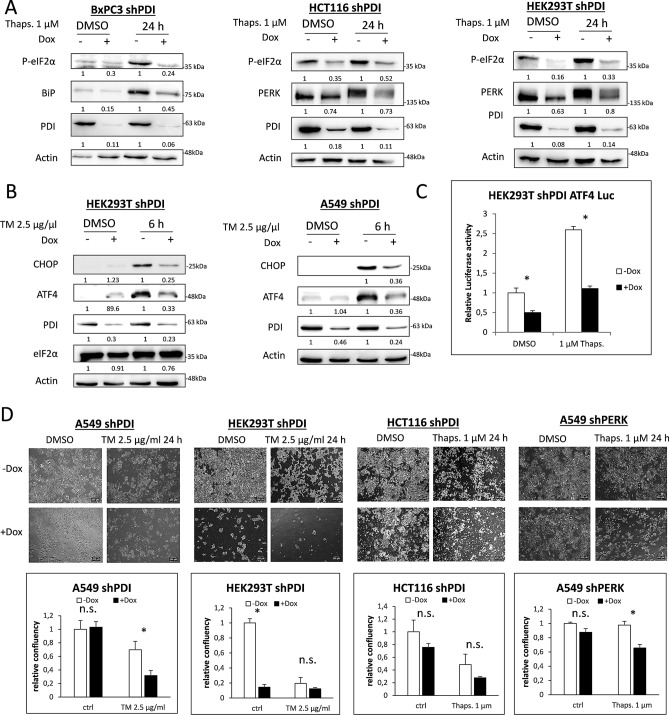
Figure 2.
PDI is essential for PERK activation after long time exposure to ER stress. (A) KD of PDI was induced for 72 h in BxPC3, HCT116 and HEK293T cells. Cells were then treated with DMSO or 1 µM thapsigargin (Thaps.) for 24 h and phosphorylation of eIF2α, expression of BiP, PERK, PDI and Actin were monitored by western blotting. (B) HEK293T and A549 shPDI cells were treated with DMSO or 2.5 µg/ml tunicamycin (TM) for 6 h in the presence or absence of PDI. Expression of CHOP, ATF4, PDI, eIF2α and Actin were tested by western blot analysis. In subfigures A and B, densitometry was used to quantify protein band intensity. After normalization to Actin, the induced KD sample (+ Dox) was compared to its control sample (− Dox, set to 1). (C) ATF4 luciferase assay in HEK293T shPDI cells after 96 h of KD induction and 20 h of 1 µM thapsigargin treatment. (D) Bright-field microscopy pictures and quantification of cell confluence of A549, HEK293T and HCT116 shPDI cells after 96 h of KD induction and 24 h treatment with DMSO, 2.5 µg/ml tunicamycin (TM) or 1 µM thapsigargin (Thaps.). Magnification × 4, 2.5 NA.

