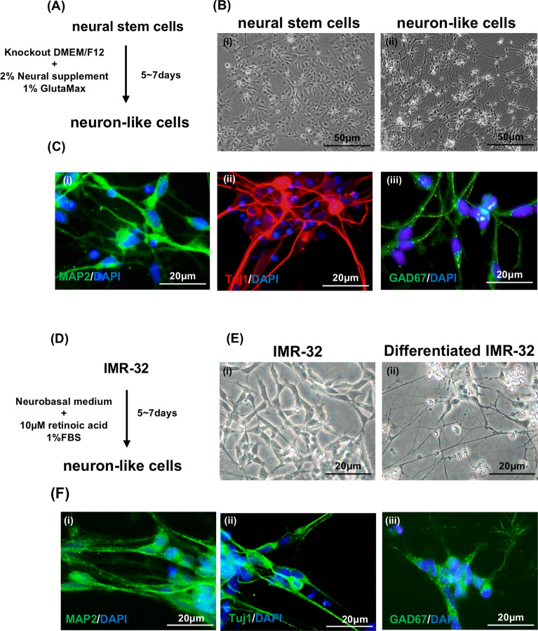
Figure 1.
The characteristics of human NSCs and neuroblastoma IMR-32-derived neuronal cells. (A) Schematic diagram of neuronal differentiation from human NSCs. (B) Bright-field image of human NSCs (i) and neurons (ii), the scale bar represents 50 μm. (C) Immunostaining images of neurons derived from human NSCs showing the expression of MAP2 (i), Tuj1 (β-tubulin III) (ii) and GAD67 (iii). The nuclei are counterstained with DAPI. The scale bar represents 20 μm. (D) Schematic diagram of neuronal differentiation from neuroblastoma IMR-32 cells. (E) Bright-field image of undifferentiated and differentiated IMR-32 cells. (F) Immunostaining images of neuronal cells derived from neuroblastoma IMR-32 cells showing the expression of MAP2 (i), Tuj1 (β-tubulin III) (ii) and GAD67 (iii). The nuclei were counterstained with DAPI. The scale bar represents 20 μm.

