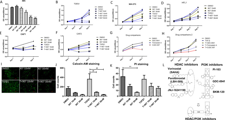
Fig. 1. CUDC-907 inhibits TGFβ1 induced fibroblasts proliferation.
The antiproliferative effect of CUDC-907 was tested in NIH-3T3 cell line, Human fetal lung fibroblasts (HLF1) and two primary cancer-associated fibroblasts (CAF1 & CAF2) with or without TGFβ1 treatment by CCK-8 assay. OD value was measured at length of 592 nm. a NIH-3T3 cells were treated with CUDC-907 in concentration gradient for 48 h. b TGFβ1-stimulated NIH-3T3 cells were treated with CUDC-907 at dose ranging from 5 to 50 nM. c–f Fibroblasts cell lines stimulated with or without TGFβ1 were treated with CUDC-907 10 nM and 30 nM for 3 days, respectively. g CAF1 were treated with 30 nM GDC-0941, Trichostatin A, and CUDC-907, respectively. h HLF1 stimulated with TGFβ1 were treated with CUDC-907. i Fluorescence microscopic images of NIH-3T3 cells stimulated with or without TGFβ1 72 h post treatment with 10/30 nM CUDC-907 and exposed to Calcein AM fluorescent dye. Scale bars: 100 µm. j Cell numbers were counted and analyzed from Calcein AM staining images. k Cell numbers were count in PI staining images (Images were not shown). Error bars are ±SD. *, **, *** represent 907 treatment groups compared with DMSO group, respectively. l Chemical structure of CUDC-907. *P < 0.05; **P < 0.01; ***P < 0.001. #, ##, ### represent T + 907 treatment groups compared with TGFβ1 group, respectively. #P < 0.05; ##P < 0.01; ###P < 0.001.

