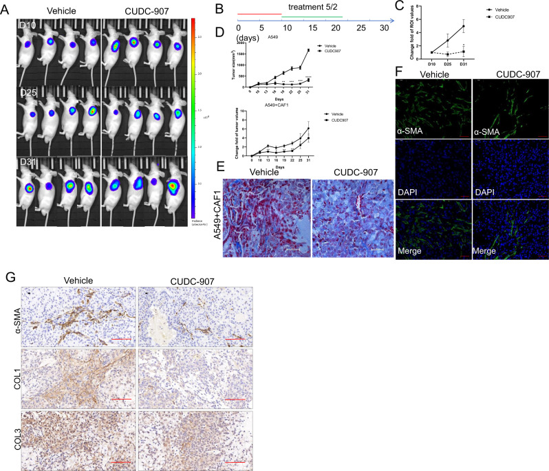
Fig. 7. CUDC-907 treatment inhibits tumor growth and fibrosis.
a Mixed A549 and CAF1 transduced with D-luciferase were injected subcutaneously into NOD/SCID mice and in vivo bioluminescent signal was quantified before and after CUDC-907 treatment. Representative in vivo images of PBS or CUDC-907 treated (5/2, 50 mg/kg) mice are acquired. b treatment schema. Mice were divided into 2 groups: Vehicle and treatment (n = 5 in each group) and injected with A549 cells or mixed A549 and CAF1(3:2). Treatment with CUDC-907 was started on day 10. CUDC-907 was administered by oral gavage (5/2, 50 mg/kg). c Tumor-to-background ratios of fluorescence intensities (n = 5). CUDC-907 treatment significantly decreased tumor fibrosis as measured by whole-body luciferase activity. d CUDC-907 treatment significantly decreases tumor growth with or without fibroblasts mixture. e Masson’s trichrome staining of tumor sections from Vehicle or CUDC-907 treated mice 10 days after subcutaneous injection of mixed A549 and CAF1. Scale bars: 100 µm. f Representative IF staining pictures in Vehicle and treated group mice. Scale bars: 25 µm. g IHC for Collagen 3 and α-SMA in Vehicle or CUDC-907 chow mice. Representative pictures are acquired. Scale bars: 100 µm. Error bars are ±SD. *, **, *** represents 907 treatment group compared with vehicle group. *P < 0.05; **P < 0.01; ***P < 0.001.

