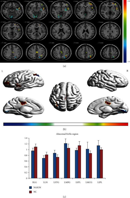
Figure 1.

Spontaneous brain activity in patients with NAION and healthy participants Significant differences in activity were observed in patients with NAION in the left middle frontal gyrus, left middle cingulate gyrus, left superior temporal gyrus, left inferior parietal lobule, right lingual gyrus, left putamen/lentiform nucleus, and left superior parietal lobule (false discovery rate corrected, cluster size > 23 voxels, P < 0.05) (a, b). The mean ReHo values for NAION and NC groups (c). Abbreviations: NAION: nonarteritic anterior ischemic optic neuropathy; NCs: normal controls; ReHo: regional homogeneity; L: left; R: right.
