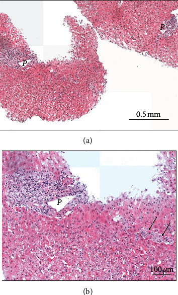
Figure 2.

Histological view of the liver biopsy specimen (hematoxylin-eosin stain) showing normal architecture and no fibrosis (original magnification ×100, panel A). At a closer view (panel B, original magnification ×200), an inflammatory infiltrate is present in the portal tract P, predominantly lymphocytic, associated with scattered eosinophils. In the liver lobule, few necrotic hepatocytes and occasional microgranulomas (arrows) are visible. Periodic acid Schiff (PAS) and Ziehl staining are negative for infections.
