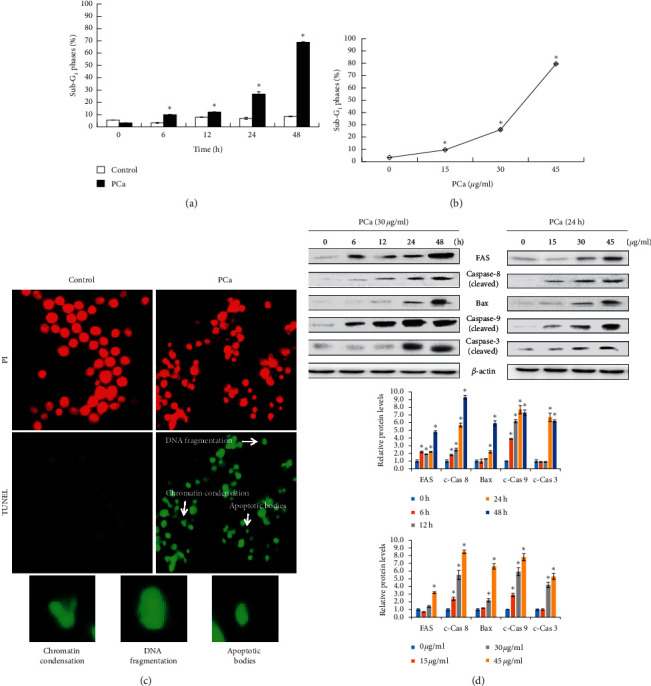
Figure 4.

PCa triggered cell apoptosis through extrinsic and intrinsic pathways in HT-29 cells. (a, b) HT-29 cells were treated with PCa and analyzed for SubG1 phase by flow cytometry. Data are expressed as mean ± SD. ∗P < 0.05 versus control group with a significant increase. (c) The typical morphology of cell apoptosis, such as chromatin condensation, DNA fragmentation, and apoptosis body, was observed after PCa treatment for 48 h by using TUNEL assays. (d) Determination of the apoptosis pathway by Western blots of PCa-treated cells.
