Figure 2.
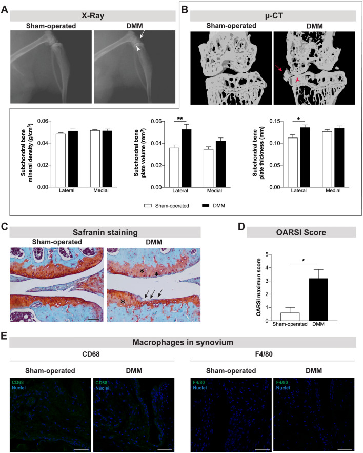
Joint damage was observed 12 weeks after the induction of the OA post-traumatic DMM model. X-ray (A) and μ-CT (B) images were acquired 12 weeks post-surgery in sham-operated and DMM mice. The narrowing of the knee joint space (arrowhead) and the formation of osteophytes (arrow) were observed in both technical approaches. μ-CT quantitative analyses were also performed (B). Increased subchondral bone volume and subchondral bone thickness were observed in the DMM mice. The safranin O staining (C) revealed intense fibrillation and thinning of the cartilage (arrows) and lower intensity in red staining (proteoglycan) of cartilage (highlighted with *) in the DMM mice (scale bar = 100 μm). A higher OARSI maximum score of articular cartilage was calculated for the DMM mice (D). The infiltration of synovium by macrophages was assessed by the analysis of CD68 and F4/80 expression and no macrophages were observed (E) (scale bar = 50 μm). Results are presented as mean ± SEM, n = 5 per group. *p < 0.05, **p < 0.01.

