Figure 6.
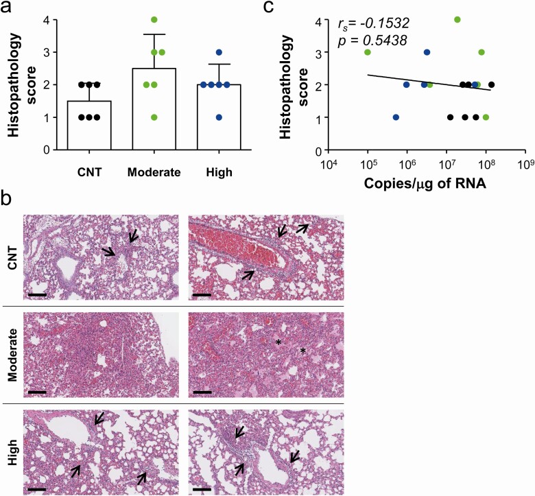
Pathological changes in lungs of hDD4-Tg mice infected with lethal dose of MERS-CoV. A and B, Lung tissue sections collected from mice at 4 days after infection were stained with hematoxylin and eosin. Pathological scores of infected lungs (n = 6/group) (bar graphs: mean + SD, A) and representative scanned images are presented (B). Bar, 100 μm. C, Correlation of histopathological scores with viral loads (copy numbers of viral RNAs) was assessed by linear regression (black line) and Spearman’s rank test (rs and P value). Abbreviations: CNT, control group; MERS-CoV, Middle East respiratory syndrome coronavirus.

