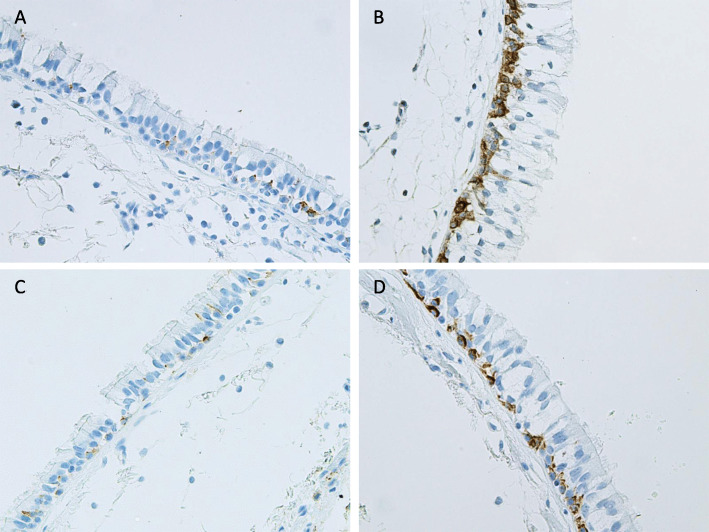
Fig. 1.
Representative images of the immunohistochemical stainings for cell-bound MUC1 in bronchial biopsy samples. Intensity of the expression was designated as negative, faint, moderate, strong or very strong, and the extent of the positive staining was estimated from 0 to 100% in each cell type present in the airways. Expression of MUC1 in cells suggesting basal cell phenotype in the large airways of a Never-smoker with normal lung functions (a), a Smoker with normal lung function (b), MUC1 expression in basal cells of an ex-smoker with COPD (c) and a Smoker with COPD (d)

