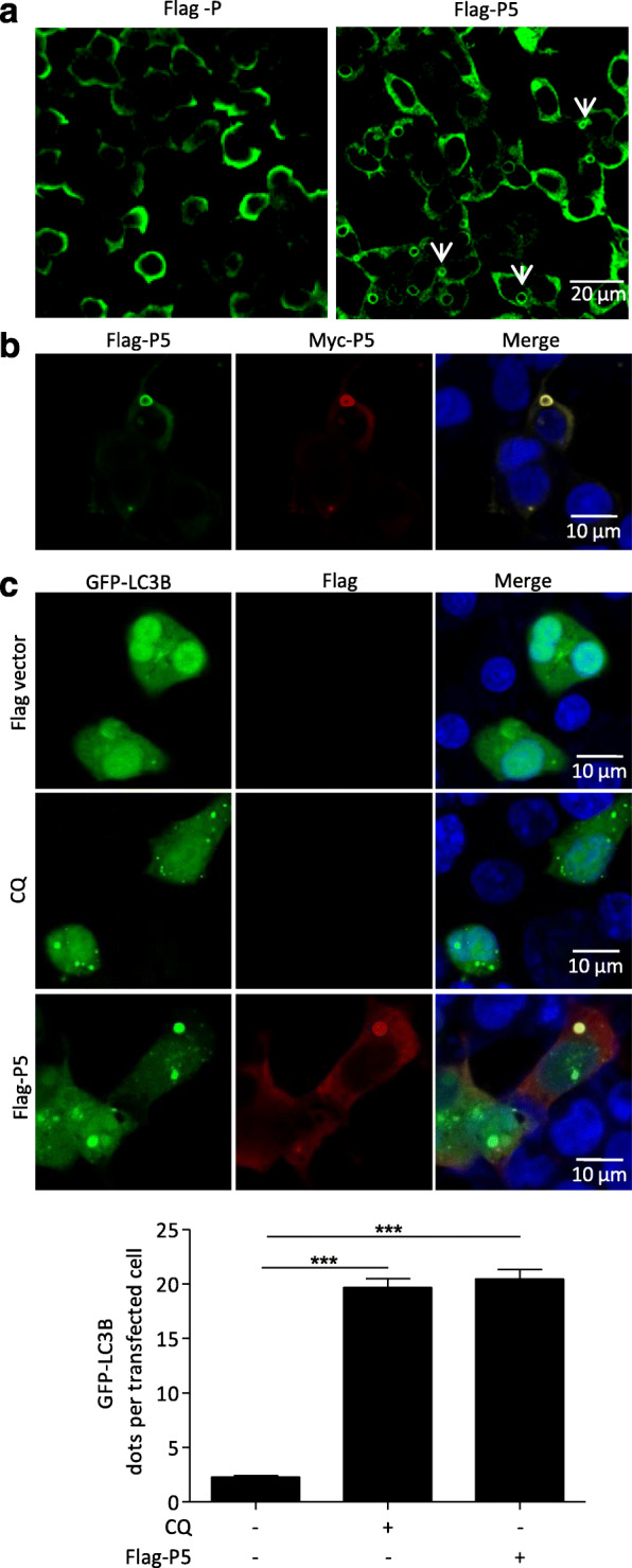
Fig. 2.

Accumulated autophagosomes are surrounded by a ring-like structure comprising P5. a-c N2a cells were transfected with Flag-P or Flag-P5, or cotransfected with Flag-P5 and Myc-P5 or GFP-LC3B plasmids for 24 h, fixed, and immunostained with mouse anti-Flag antibodies and rabbit anti-Myc antibodies, and then visualized using confocal microscopy. DAPI (blue) was used to stain nuclear DNA. Scale bar: 10 or 20 μm. The graph shows the quantification of autophagosomes by taking the average number of dots in 50 cells. Means and SD (error bars) of three independent experiments are indicated (*, P < 0.05; **, P < 0.01; ***, P < 0.001)
