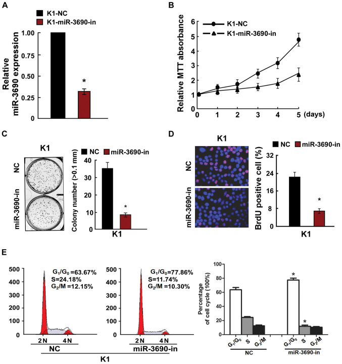
Figure 3.
Inhibition of miR-3690 inhibited TC cell proliferation. (A) Validation of miR-3690 expression levels after transfection by PCR analysis. (B) MTT assays revealed that miR-3690-in suppressed growth of K1 cells. (C) Inhibition of miR-3690 inhibited the anchorage-independent growth of K1 cells. Representative micrographs (left) and quantification of colonies that were >0.1 mm (right). (D) Representative micrographs (left) and quantification (right) of the BrdU incorporation assay in K1 cells. (E) Flow cytometric analysis of the indicated TC cell line K1 cells transfected with NC or miR-3690-in. Each bar represents the mean of three independent experiments. *P<0.05 vs. NC group. miR, microRNA; TC, thyroid cancer; NC, negative control; miR-3690-in, microRNA-3690-inhibitor.

