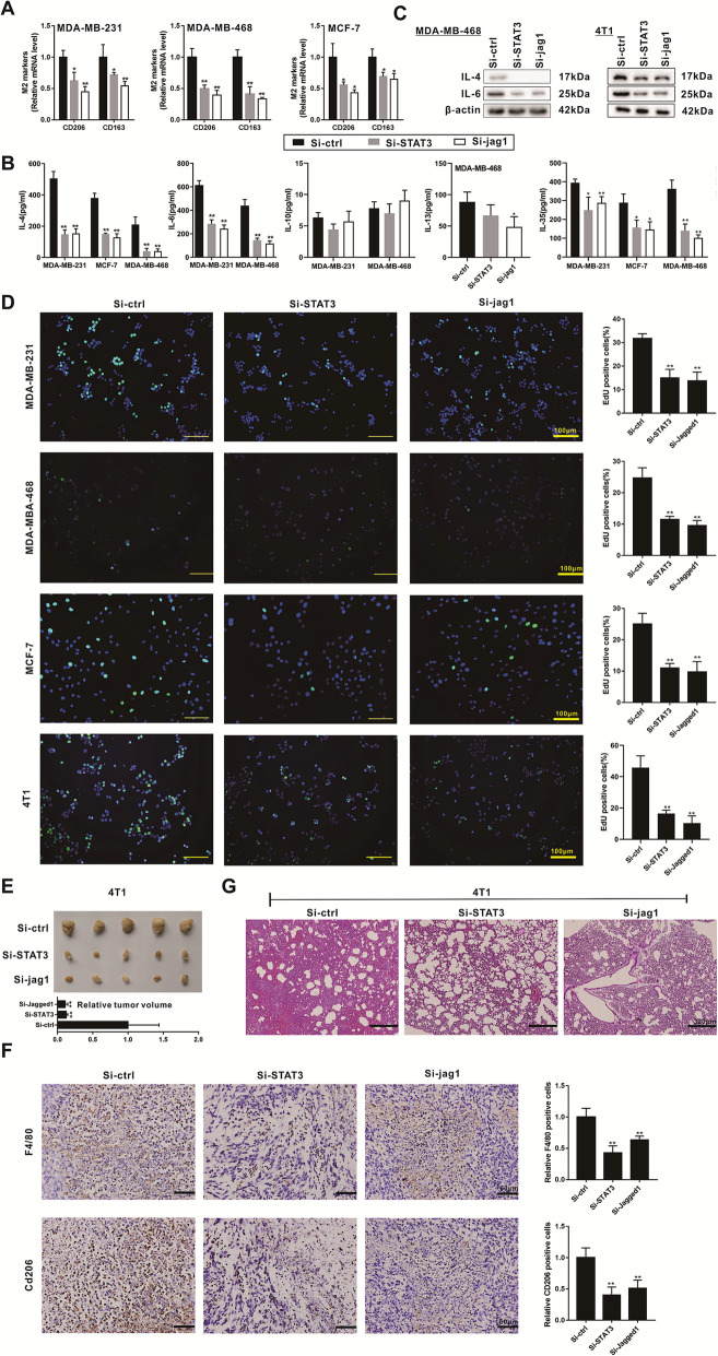
Fig. 4.
STAT3 and Jagged1 participates in the breast cancer metastasis and M2 polarization of macrophages. a. Human breast cancer cell lines (MDA-MB-231, MDA-MB-468, and MCF-7) were transiently transfected with STAT3 siRNAs (Si-STAT3) or JAG1 siRNAs (Si-JAG1) for 48 h before the co-culture. After co-culturing breast cancer cells with PMA-induced human monocyte THP-1 cells for 48 h using Transwell assay, we detected the relative mRNA levels of CD206 and CD163 in THP-1 derived macrophages, both of which were M2 polarization markers of macrophages. b-d. Human breast cancer cell lines (MDA-MB-231, MDA-MB-468, and MCF-7) and mouse breast cancer cell line (4 T1) were transiently transfected with STAT3 siRNAs (Si-STAT3) or JAG1 siRNAs (Si-JAG1) for 48 h. The level of IL-4, IL-6, IL-10, IL-13, and IL-35 in supernatants was detected using ELISA (b). The protein level of IL-4 and IL-6 in breast cells was detected using western blot analysis (c). The cell proliferation was detected using EdU labeling detection (d). Scale Bar =100 μm. e-f. Female Balb/c mice were subcutaneously injected with the mouse breast cancer cell line (4 T1, 5 × 10⁵ cells) which were transfected with STAT3 siRNAs (Si-STAT3), JAG1 siRNAs (Si-JAG1), or the control siRNAs (Si-ctrl) (n = 10 in each group). The tumor tissues were collected and measured on the 20th day (e). The F4/80 and CD206 expressions in tumor tissues were detected using immunohistochemistry (f). Scale bar = 60 μm. g. Female Balb/c mice were intravenously injected with the 4 T1 cells (1.5 × 10⁵ cells) which were transfected with STAT3 siRNAs (Si-STAT3), JAG1 siRNAs (Si-JAG1), or the control siRNAs (Si-ctrl) (n = 5 in each group). The pulmonary metastatic tumor tissues were observed using HE staining. Scale Bar = 320 μm. Three independent experiments. *P < 0.05, **P < 0.01 vs control (Si-ctrl)

