Figure 3.

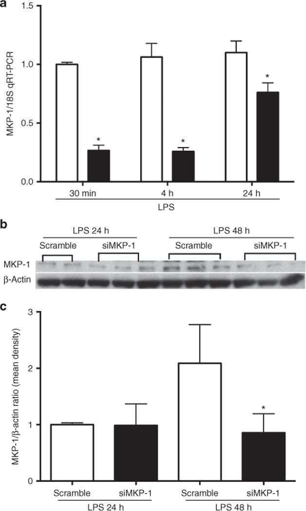
MKP-1 knockdown in rIEC-6 cells. (a) rIEC-6 cells were transiently transfected with either siRNA against MKP-1 (siMKP-1; black bars) or scramble siRNA (scramble; white bars). Twenty-four hours after transfection, the cells were stimulated with LPS (100 μg/ml) for the indicated times. MKP-1 expression was evaluated by qPCR following transfection; *P < 0.05 vs. scramble for each time point. (b) Representative western blot of MKP-1 protein expression after transfection with siRNA for MKP-1 or scramble siRNA. Following transfection, rIEC cells were stimulated with LPS for 24 and 48 h. (c) Densitometry data shown as means ± SEM of protein level relative to β-actin protein levels for triplicate samples from three independent experiments. *Protein expression of MKP-1 after siMKP-1 transfection and LPS stimulation for 48 h is different from MKP-1 protein expression after scramble transfection and LPS stimulation for 48 h, P < 0.05.
