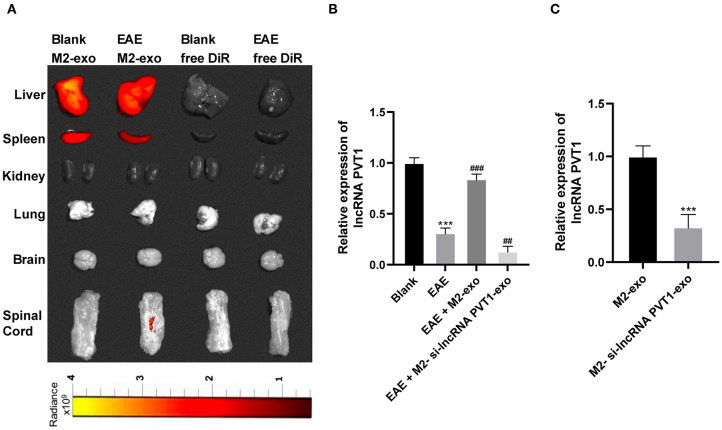
Figure 3.
LncRNA PVT1 enters EAE mice from M2-exos. (A) DiR labeled exosomes were traced in mice. It showed that exosomes mainly existed in liver and spleen 24 h after they entered the mice. Fluorescence also appeared in spinal cord of EAE mice; (B,C) Relative PVT1 expression in spinal cord of EAE mice with different treatment detected by RT-qPCR. Compared with the blank group, ***p < 0.001; compared with the EAE group, ##p < 0.01, ###p < 0.001. Data in (B) are analyzed with one-way ANOVA, and Tukey's multiple comparisons test was utilized for post hoc test. Data in (C) were analyzed by the t-test n = 6.

