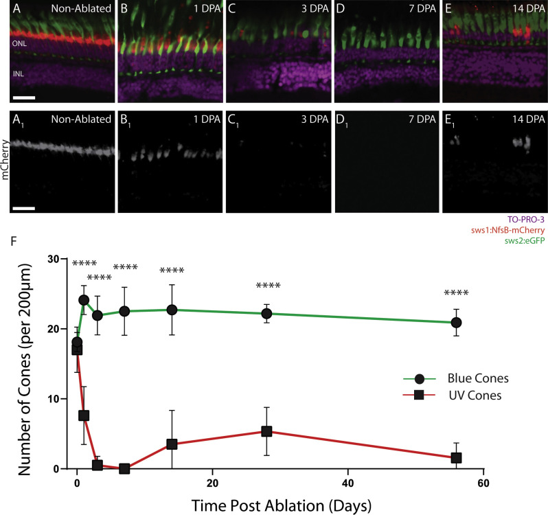
Figure 1.
Retinal histology shows loss and regeneration of UV Cones after metronidazole treatment in sws1:nfsB-mCherry;sws2:eGFP zebrafish. Non-cone-ablated fish (A) show UV (sws1) and blue (sws2) cones with regular spacing and morphology. At 1 DPA (B), the outer segments of UV and blue cone show blebbing and elongation, but the pedicles have only retracted in the UV cones. At 3 DPA (C), the blue cones seem to have regained normal outer segment morphology, and only remnants of the UV cones remain. At 7 DPA, no UV cones were observed using histology (D). UV cones are observed to have regenerated by 14 DPA (E), but at a reduced number compared to the preablated UV cone density. By 3 DPA, UV cones have dramatically decreased, and there are significantly fewer UV cones (F). Scale bar: 25 µm. ONL, outer nuclear layer; INL, inner nuclear layer. *P < 0.05; **P < 0.01; ***P < 0.001; ****P < 0.0001.

