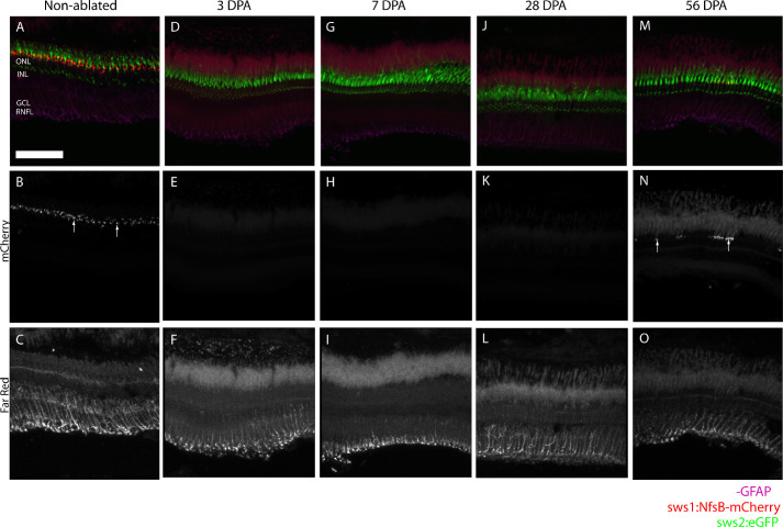
Figure 2.
Histology shows no increase in GFAP staining and Müller cell activation after metronidazole treatment. Untreated zebrafish (A) show a continuous UV cone mosaic (B, arrows) and baseline Müller cell activation (C). At 3 DPA (D), UV cones aren't observed (E) and no increased in Müller cell activation is seen (F). Similarly, at 7 DPA and 28 DPA (G,J), histology shows no UV cones (H, K) and no Müller cell activation (I, L). At 56 DPA (M), some UV cone regeneration is observed (N, arrows), though the UV cone mosaic is not regular. Despite the UV cone regeneration, no increased in Müller cell activation is observed from GFAP staining (O). Scale bar: 100 µm.

