Figure 3.
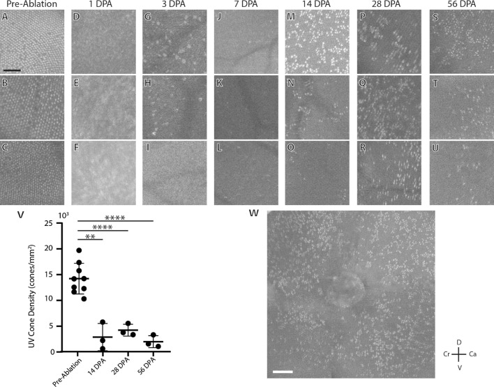
Cross-sectional variability in the UV cone mosaic. Each image shown is a retina from separate fish, except for panel W, which shows a larger montage of panel M. Pre-cone-ablated zebrafish (A, B, C) show a regular UV cone mosaic. At 1 DPA (D–F), no cones are observed in any of the fish. At 3 DPA (G–I), cones are still not observed, but large, hyperreflective inclusions can be observed in some fish (G, H). Individual UV cones are observed at 7 DPA (J–L) and by 14 DPA (M–O) regenerated UV cones show a wide range of cone densities and an irregular clumping pattern. Irregular reflective cones are still observed at 28 DPA (P–R) and 56 DPA (S–U). UV cone density measured from OCT is significantly decreased at 14, 28, and 56 DPA (V). A montage of a fish 14 DPA shows that UV cone regeneration does not follow a gradient and occurs equally across the retina (W). D, dorsal; V, ventral; Cr, cranial; Ca, caudal. Scale bars: 50 µm (A–U) and 100 µm (W). *P < 0.05; **P < 0.01; ***P < 0.001; ****P < 0.0001.

