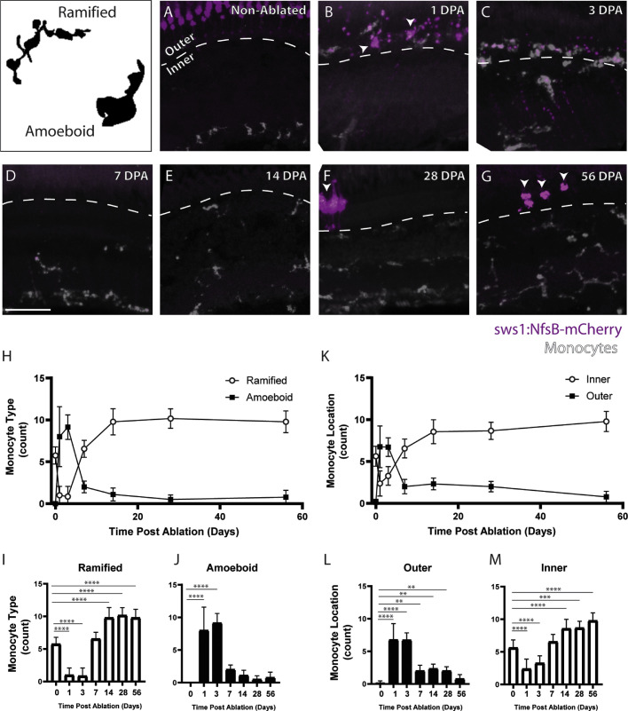
Figure 6.
Histology shows a change in monocyte location and morphology after metronidazole-mediated ablation of UV cones. In nonablated retinas (A), monocytes are observed to have a largely ramified morphology and to be distributed largely throughout the inner retinal layers and UV cones appear to have a normal morphology. At 1 DPA, monocytes have largely switched to an amoeboid appearance and translocated to the outer retina (dashed line, B); the cones at 1 DPA have begun to form blebs and lack the typical cone morphology (B, arrows). Monocytes at 3 DPA continue to have a largely amoeboid appearance and to be in the outer retinal layers (C). The cones at 3 DPA have been largely cleared, and only a few small blebs remain. By 7 DPA, the cones have been completely cleared, and the monocytes have returned to the inner retina with a ramified morphology (D) and remain in this state at 14 DPA (E), 28 DPA (F), and 56 DPA (G). Regenerated UV cones are observed at 28 and 56 days after ablation (F, G, arrows). Graphs of monocyte morphology (H–J) and location (K–M) show the trends observed in histology. Examples of amoeboid and ramified morphology are shown in the top left panel. Scale bar: 50 µm. *P < 0.05; **P < 0.01; ***P < 0.001; ****P < 0.0001.

