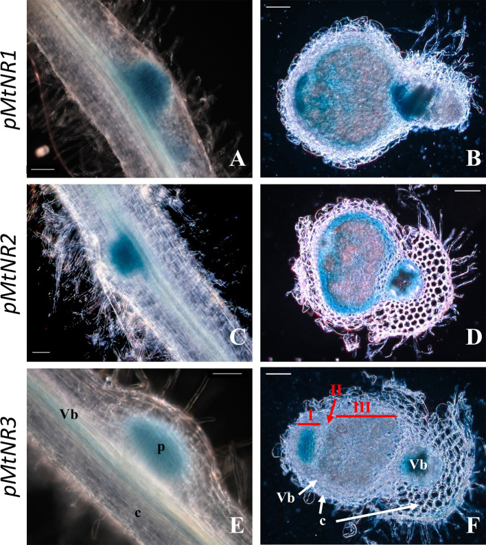
Figure 2.
Histochemical localization of MtNRs expression in Medicago truncatula roots. Localization of GUS activity in transgenic M. truncatula roots expressing the gusA reporter gene under the control of a 1.65 Kb MtNR1 promoter fragment (A, B), of a 1.7 kb MtNR2 promoter (C, D) and of a 1.55 kb pMtNR3 promoter (E, F). Whole root segment 4 dpi with S. meliloti (A, C, E) and longitudinal section of a 2 wpi old nodules (B, D, F) were stained for 3, 5, or 16 h with X-gluc for the GUS activity for pMtNR2, pMtNR1, or pMtNR3 respectively. Zones I, II, and III of the nodule are represented in red in picture F. p, nodule primordium; Vb, vascular bundles; c, cortex. Scale bars, 50 µm for (A, C, E); 100 µm for (B, D, F).

