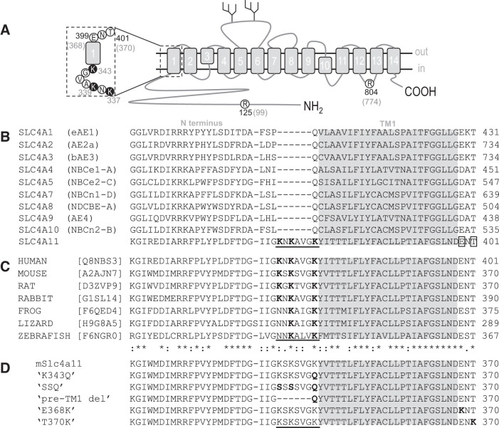
Fig. 6.
Slc4a11 residues mutated in this study. A: topological cartoon of Slc4a11 showing the 14 transmembrane spanning regions. Amino-acid residues pertinent to this study are circled and numbered according to their location in the human (black numbers) or mouse (gray numbers) polypeptide chain. The region that corresponds to the sequence alignments in B–D is emphasized. B: alignment of all human SLC4 family protein sequences showing a unique lysine-rich region in SLC4A11 (underlined) and 2 nearby residues mutated in corneal dystrophy (E399K and T401K, boxed). Gray region shows likely limits of the first transmembrane span C: alignment of a selection of vertebrate Slc4a11 sequences showing conservation of the sequence features highlighted in Fig. 5B. D: sequence of the mutants used in this study (altered regions are shown in bold).

