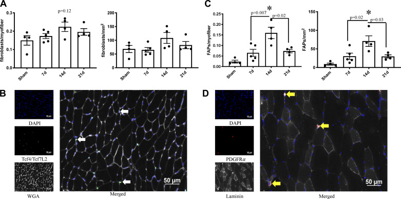
Fig. 2.
Fibrogenic cell abundance is elevated in skeletal muscle after severe burn injury. A: fibroblast abundance relative to myofiber number and area of spinotrapezius cross section. B: representative images of fibroblast abundance in spinotrapezius cross section. Tcf4/Tcf7L2 and DAPI costaining denote myofibroblasts (white arrow). C: fibrogenic adipogenic progenitor cell (FAP) abundance relative to myofiber number and area of spinotrapezius cross section. D: representative images of FAP abundance in spinotrapezius cross section. Platelet-derived growth factor receptor-α (PDGFRα) and DAPI costaining denote FAPs (yellow arrow). Data are presented as means ± SE. One-way ANOVA with Tukey’s post hoc test. *P < 0.05 compared with sham; n = 4–5 mice/group.

