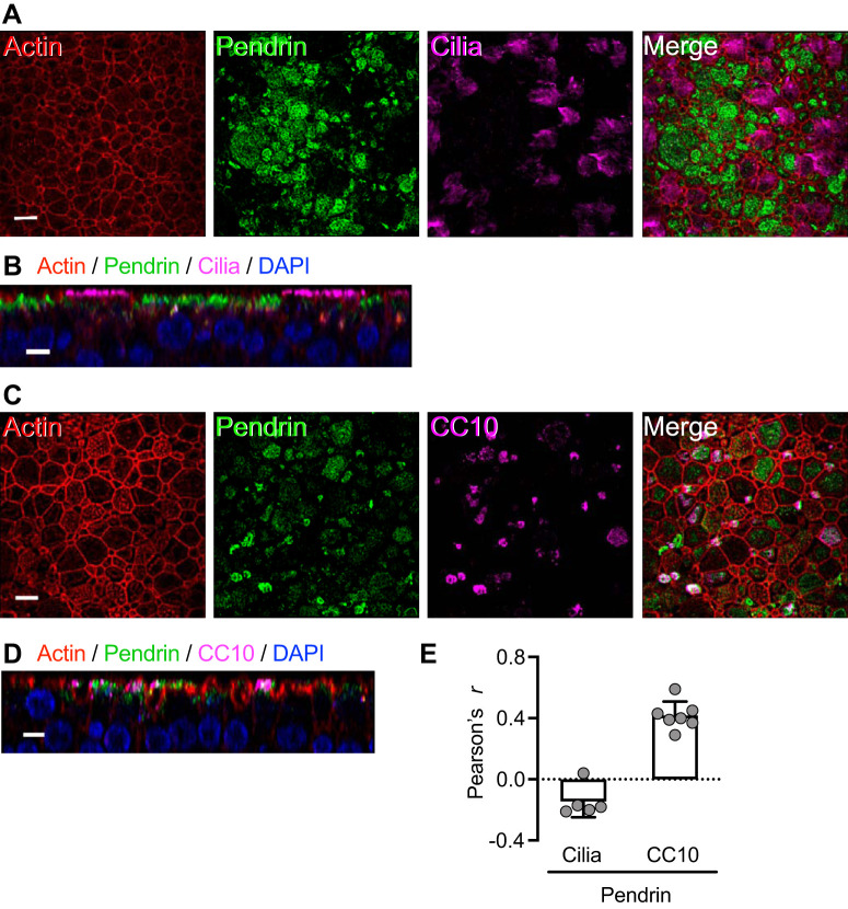
Fig. 9.
TNFα+IL-17 induce pendrin expression mainly in secretory cells. Human airway epithelia were treated with TNFα+IL-17 for 48 h. A and B: ciliated cells lacked significant pendrin expression. A shows an en face projection, and B shows an X-Z projection. C and D: pendrin-expressing cells were, in many cases, CC10+, which labels secretory cells. C shows an en face projection, and D shows an X-Z projection. Scale bars: A and C, 10 μm; B and D, 5 μm. E: colocalization of pendrin with ciliated cell and CC10 markers measured using the ImageJ software and reported as Pearson’s correlation coefficient r (n = 5–7).

