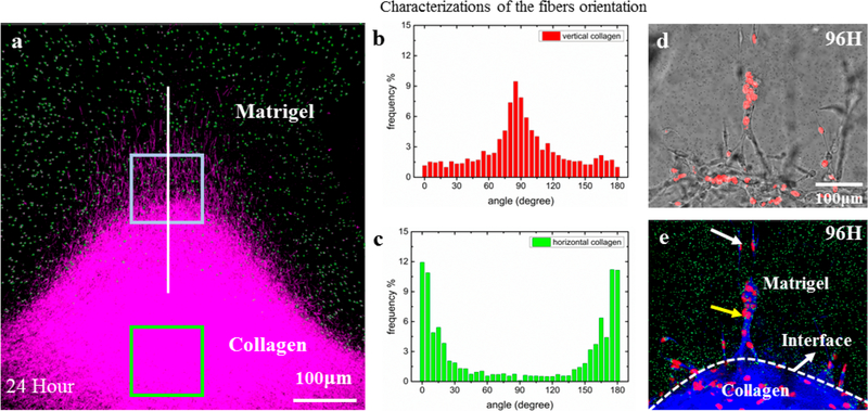
Figure 5.
Oriented collagen fibers direct the metastatic cell invasion into Matrigel. (a) The 3D confocal image reconstruction via image stacking (top view) shows the Matrigel/collagen composite ECM and their interface in three dimensions. (b) The collagen fibers near the interface region possess vertical orientations (red box). (c) The collagen fibers in the centered region possess horizontal orientations (green box). (d) The bright-field images showing snapshots of invading cells at 96 h. (e) The corresponding fluorescent images combined with reflective mode of panel d, which show the Matrigel region with green beads embedded, the collagen region (blue), and the nuclei of invading cells (red). It is clear that, at the 96th hour, guided by oriented collagen fibers, the cells aggregated and strongly invaded into the rigid Matrigel region in single-stream forms. The field of view is the same for panels d and e. Adapted with permission from ref 127. Copyright 2016 Weijing Hana, Shaohua Chen, Wei Yuan, Qihui Fana, Jianxiang Tian, Xiaochen Wang, Longqing Chen, Xixiang Zhang, Weili Wei, Ruchuan Liu, Junle Qu, Yang Jiao, Robert H. Austin, and Liyu Liu.

