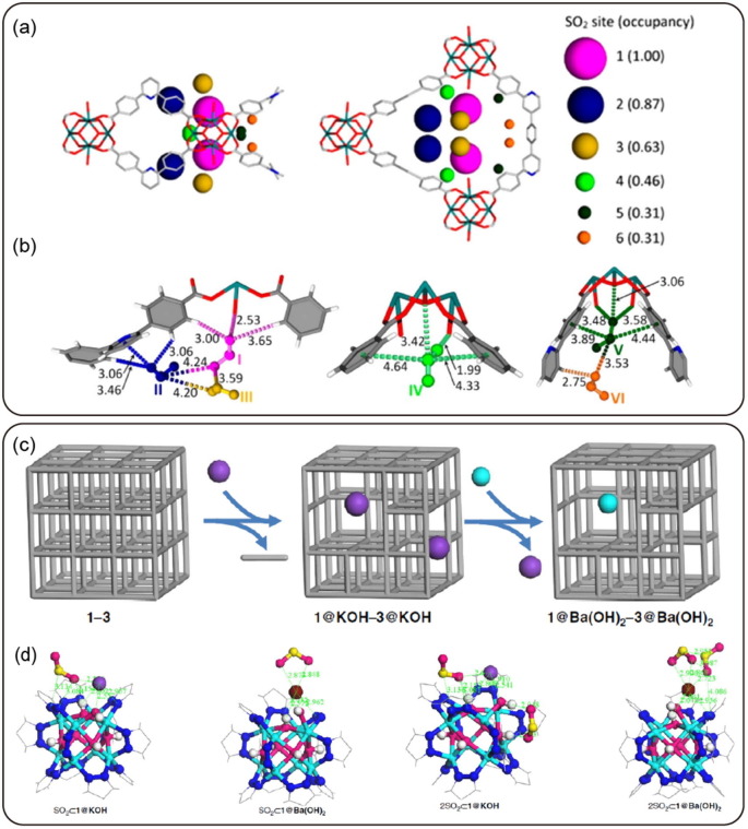
Fig. 31.
(a) Site occupancy (illustrated by spheres) of SO2. (b) Binding sites of SO2 in MFM-601 as refined by in-situ synchrotron PXRD. Reproduced with permission from [249]. Copyright 2018 American Chemical Society. (c) Schematic illustration of the successive PSMs between potassium (purple) and barium (cyan) from pristine nickel pyrazolate. (d) The DFT structure of sulfur dioxide interaction with crystal defect sites. Reproduced with permission from [252]. Copyright 2017 Springer Nature.

