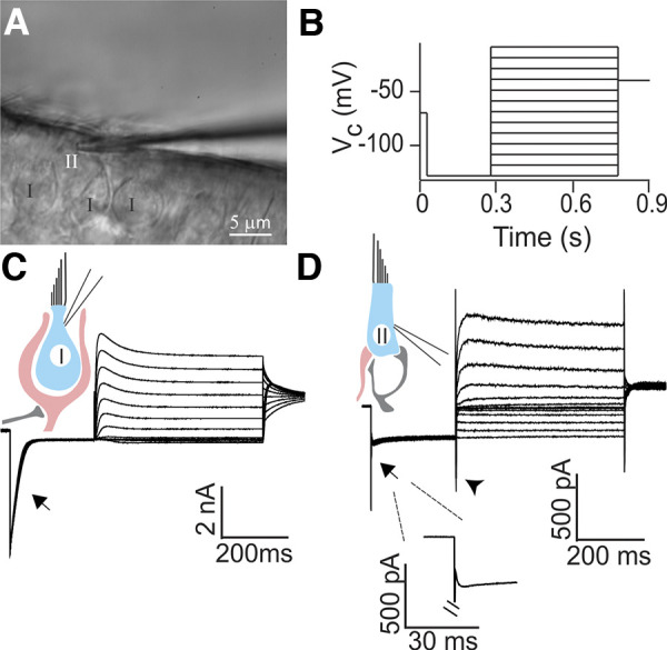
Fig. 1.

Whole cell patch-clamp recording from morphologically and physiologically identified rat type II hair cells (HCs). A: in a whole mount preparation of the rat crista, putative type II HCs were recognized based on the lack of a surrounding calyx using differential interference contrast (DIC) optics. Electrode is shown approaching the HC from the right. B: characteristic conductances of HCs were examined by a voltage-clamp protocol consisting of a 250-ms hyperpolarization step from −70 mV to −130 mV (held for 250 ms) followed by a family of 500-ms depolarization steps with an increment of 10 mV up to a holding potential of −10 mV. A final potential of −40 mV was held for 100 ms. Vc, voltage command. C: type I HCs showed a large, slowly inactivating inward current (IK,L; arrow) in response to a negative voltage step. D: type II HCs lacked the large IK,L current found in type I HCs (arrow, and enlarged in inset). An inward current (likely a hyperpolarization-activated current, Ih) could be seen in response to a depolarizing voltage step in type II HCs (arrowhead) as described previously (Brichta et al. 2002; Meredith et al. 2012; Rüsch et al. 1998).
