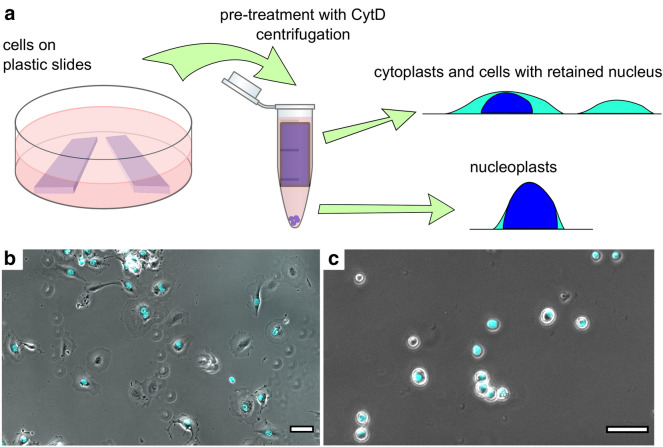
Fig. 1.
The scheme of the enucleation protocol. a Cells growing on plastic slides were inserted in 1.5 mL Eppendorf tubes filled with the cell medium and pre-treated with CytD for 15 min. After the centrifugation, the remaining cells and cytoplasts were reseeded from the plastic slides to fibronectin-coated Petri dishes, and AFM experiments were performed 3 to 5 h after. The pellet containing nucleoplasts was resuspended in HBSS and placed on poly-L-lysine-coated dishes for further experiments. b A sample of REF52 enucleated cells contained both cells without the nucleus (enucleated cells, cytoplasts) and cells with the retained nucleus. The cell-permeable DNA dye (cyan) was used to distinguish cytoplasts from normal cells. c Nucleoplasts. DNA staining was used to distinguish nucleoplasts from the cell debris. Scale bars are 50 µm

