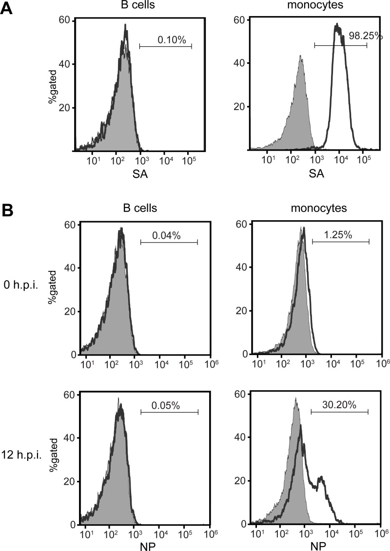
Fig 1. High expression of α2,3 SA is detected on monocyte but not on B cell surface.
Sialic acid receptors were detected on isolated B cells and monocytes by lectin staining with MAL I. B-cells and monocytes are identified by gating the CD20- and CD14-positive cells, respectively. (A) Flow cytometry analysis shows the percentage of B cells (left panel) and monocytes (right panel) positive for surface α2,3 SA (open histograms) relative to the unstained cells (grey histogram). B cells and monocytes were infected with H5N1 virus at MOI of 1 for 12 hours and subjected to flow cytometry after NP staining. (B) NP expression on B cells and monocytes at 0 and 12 hours post infection (h.p.i.) are shown (H5N1 infection, open histogram; mock infection, grey histogram). Histograms are representative of three independent experiments.

