
FIGURE 2.
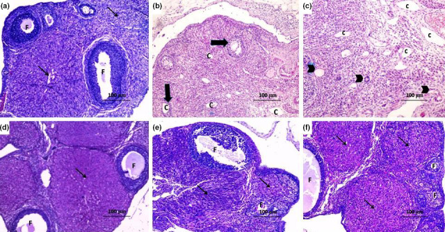
Sections of the ovary (H&E x100) from (a) the control group showing normal ovarian follicles (F) and normal corpus luteum (thin arrow); (b) DHEA group showing numerous subcapsular cysts, with a very thin granulosa layer (thick arrow), absent corpora lutea, many fluid‐filled cysts (C) with severe inflammatory cellular infiltration; (c) DHEA + CMC group showing numerous subcapsular cysts (C) with absent corpora lutea and atretic follicles containing fluid‐filled antrum (arrow head) with marked inflammatory cellular infiltration; (d) DHEA + Telmisartan group showing some healthy follicles (F) and corpora lutea (thin arrow) with disappearance of the cysts and significant decrease in the inflammatory cellular infiltration; (e) DHEA + Fish oil group showing healthy follicles (F) and some corpora lutea (thin arrow); and (f) DHEA + Telmisartan +Fish oil group showing a structure relatively near to normal with many corpora lutea (thin arrow) and antral follicles (F) with clearly differentiated oocytes, corona radiate, granulosa cell layer, and thecal cells