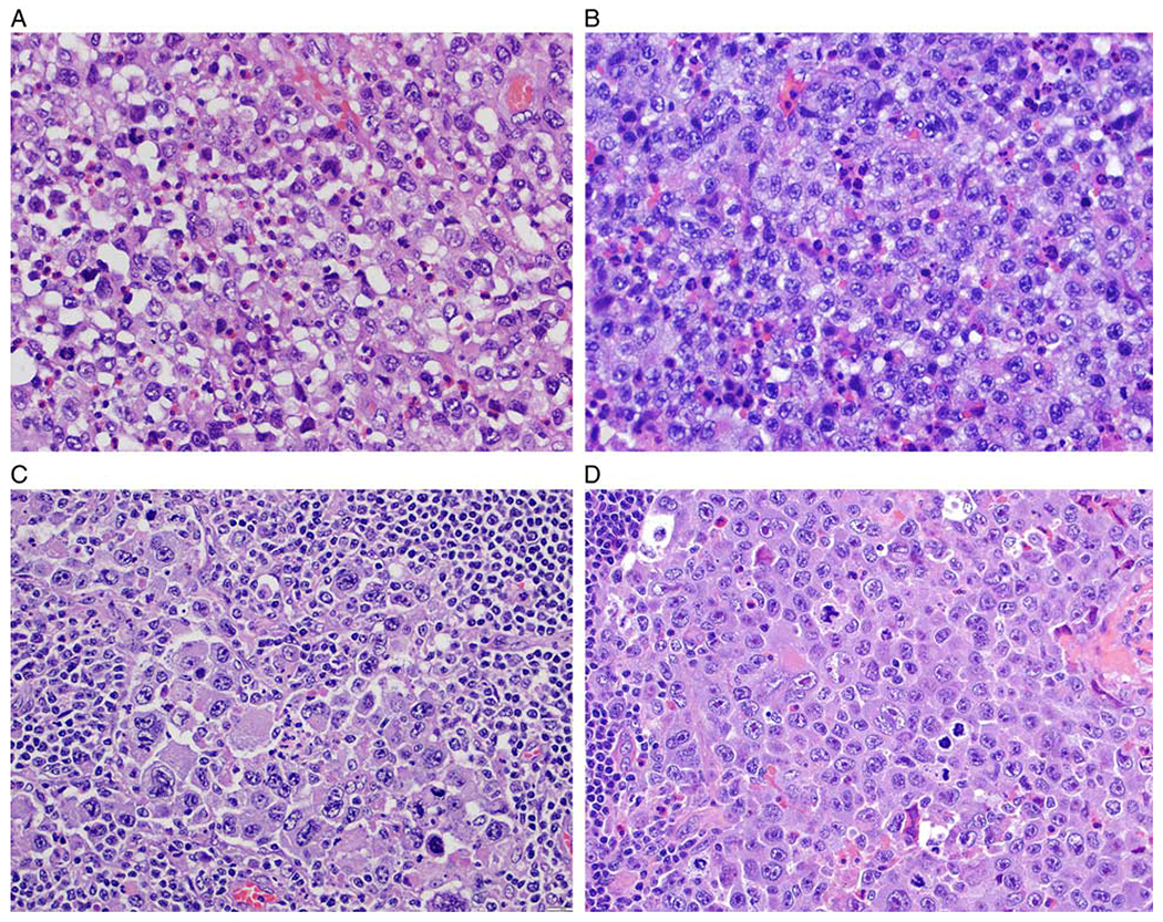
FIGURE 4.

BI-ALCL cytomorphology. A, This case displays a predominance of cells with round to oval nuclei, vesicular chromatin and distinct nucleoli admixed with scattered eosinophils. B, This case displays a predominance of cells with lobulated nuclei, vesicular chromatin and irregular nuclear membrane with indentations. C, Most of the neoplastic cells are large and pleomorphic and have vesicular or hyperchromatic nuclei. D, This case illustrates a subset of lymphoma cells with cytomorphology of “hallmark cells” with nuclear indentations, abundant cytoplasm, and distinct paranuclear clearing (all figures stained with hematoxylin and eosin).
