
Fig. 1: ZIKV-E ubiquitination on K38 and K281 promotes virus replication in cells and in vivo.
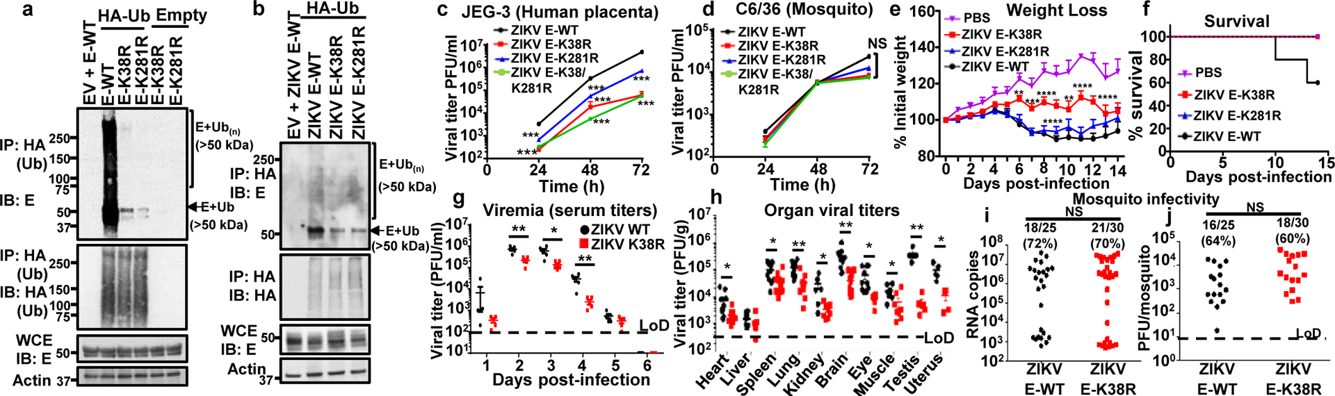
a, Whole cell extracts (WCE) from HEK293T cells transfected with empty vector (EV), E-WT or mutants and HA-Ub were used for IP with anti-HA beads. b, JEG-3 cells stably expressing HA-Ub were infected with ZIKV E-WT, or ZIKV mutants followed by AHA IP. Since the mutant viruses are attenuated, the input E was normalized for immunoprecipitation. c-d, Virus titers in supernatants from infected JEG-3 cells (c); or mosquito C6/36 (d), at MOI 0.5. Representatives from 2 independent experiments (n=3 technical replicates, mean +/−SE, ***P < 0.001). e-h, A129 mice (5 mock) or infected with ZIKV mutants (1×10⁴ PFU, 9 mice/group, from 2 independent experiments). e, Body weight (Two-way ANOVA, Tukey’s test). f, Survival. Virus titers shown in Extended Data Fig 3c–d. g, Same experiment as in e repeated with 5 males, 5 females (Viremia), and h, virus titers (day 6 p.i.). Unpaired two-sided t-test, *p<0.05, **p<0.01. i-j, Mosquito infectivity. Aedes aegypti mosquitoes were fed with bloodmeal (106 PFU/ml) of ZIKV. At day 10, individual mosquitoes were quantified for viral RNA (qPCR, i) and virus (plaque assay, j). LoD, limits of detection. Unpaired, two-sided t-test, NS (Not significant, p>0.05).