
Figure 5). Cx43 protein occurs in reduced levels at the endoplasmic reticulum and is primarily junctional during adenovirus infection.
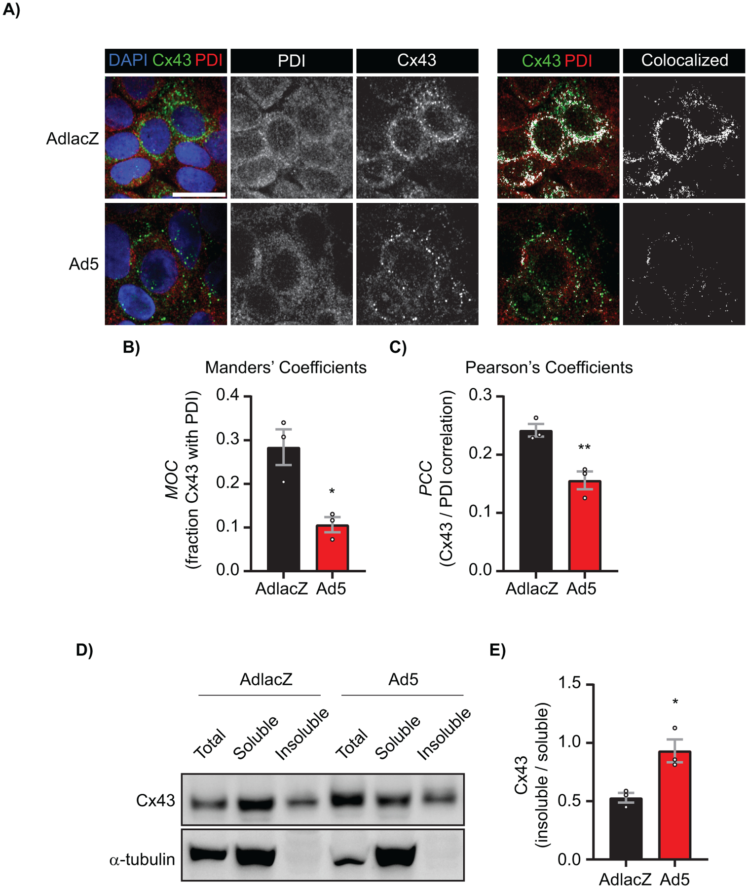
HaCaT cells were infected with AdlacZ or Ad5 at a MOI of 10 iu/cell prior to fixation or protein harvesting at 24 hpi. A) AdlacZ- or Ad5-infected HaCaT cells were immunolabeled against PDI (red) to localize ER and against Cx43 (green). Cells were stained using DAPI to identify nuclei (blue). Colocalized Cx43 / PDI signal was determined with ImageJ (white). Original magnification: X100. Scale bar: 20 μm. B) Manders’ Coefficients calculations determined using fraction Cx43 with PDI. Data points represent averages of 8 images from 3 separate experiments. C) Pearson’s Coefficients calculations determined for Cx43 and PDI correlation. Data points represent averages of 8 images from 3 separate experiments. D) AdlacZ- or Ad5-infected HaCaT cells were lysed in 1% Triton X-100 solubility assay buffer prior to fractionation and western blotting. Membrane probed against Cx43 (top panel) and α-tubulin (bottom panel) for loading control. E) Quantification of D. Statistical analysis was performed with Student’s t-test. (n=3). *p≤0.05, **p≤0.01. Data are represented as mean ±SEM.