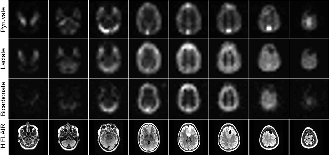
Figure 3. Example dynamic HP-13C EPI: brain data.
HP [1-13C]pyruvate, [1-13C]lactate, and [13C]bicarbonate area-under-the-curve (AUC) EPI images from eight slices covering the entire brain of a patient who has undergone treatment for brain cancerImages are devoid of Nyquist ghost artifacts or apparent geometric distortionFor anatomic reference, 1H FLAIR images are provided in the bottom row.

