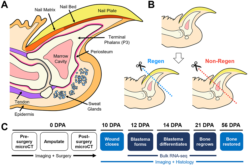
Figure 1. Schematic of the murine distal digit model.

(A) Mid-sagittal section of the mouse digit tip with major anatomical structures. (B) Level-dependent amputation of the terminal phalanx bone (P3). Blue and red dashed lines indicate amputation planes for the Regenerative (Regen) and Non-Regenerative (Non-Regen) groups, respectively. (C) Experimental schematic show time course of regeneration at various days post-amputation (DPA).
