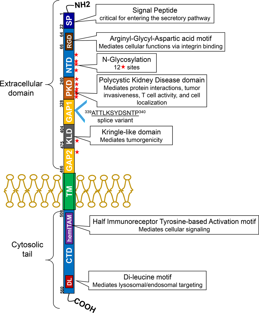
Figure 1. Structure of GPNMB.
This membrane bound protein contains a signaling domain (SP), an RGD motif in the N-terminal domain (NTD), a polycystic kidney disease (PKD) domain, and a Kringle-like domain (KLD) in the extracellular portion. The cytosolic domain, which is separated by the transmembrane domain (TM), consists of a half immunoreceptor tyrosine-based activation motif (hemITAM), and a lysosomal/endosomal targeting dileucine motif (DL) in the C-terminal domain (CTD). Solid stars indicate potential N-glycosylation sites. A GPNMB isoform is identified where 12 amino acids are inserted between amino acid 339 and 340 in GAP1, most likely due to alternative splicing.

