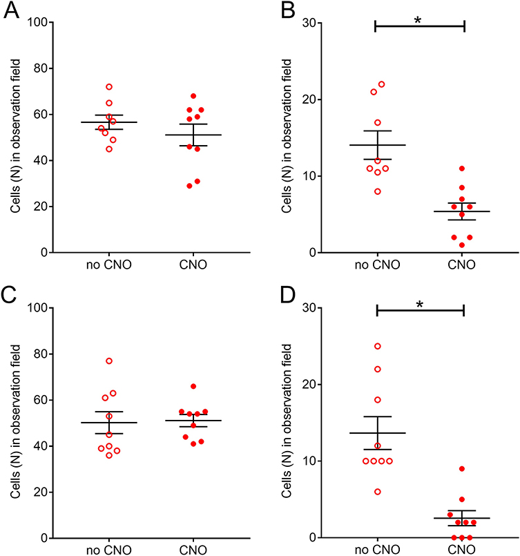
Figure 7.
Number of cells positive for pERK within the reticular thalamic nucleus (Rt). Panels A and B are for female rats and panels C and D are for males. Panels A and C are for the number of pERK positive cells in the Rt. Panels B and C are the number of cells co-localizing for both pERK and GAD67 in the Rt. Animals were injected with vehicle (no CNO) or CNO (clozapine N-oxide). Asterisk indicates a significant difference (p<0.05) between groups, there were 8 or 9 animals per group.

