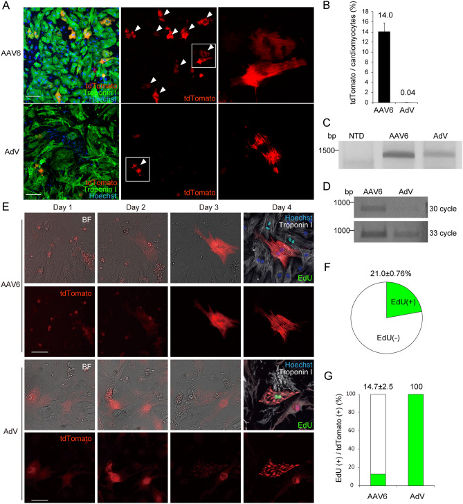
Figure 3.
Imaging-based evaluation of targeted integration in cardiomyocytes transduced with AAV or AdV. (A) Neonatal cardiomyocytes isolated from Cas9 knock-in mice were seeded in 96-well plates and transduced with AAV6 (8.3 × 10⁴ viral genomes/cell) or AdV (1.4 × 10¹⁰ PFU/ml, 1 μl/well). Five days after transduction, cells were fixed and stained with anti-troponin I antibody (green). Cardiomyocytes in which tdTomato signal co-localized with sarcomeres are indicated with arrowheads. Scale bar: 100 μm. (B) The proportion of tdTomato-positive cardiomyocytes was calculated using high-content image analysis (AAV6, n = 6, AdV, n = 8, means ± SD). (C) Cardiomyocytes were treated as in (A). Four days after transduction, genomic DNA was extracted and the targeted sequence was amplified by PCR using the primers indicated in Fig. 1A. Original full image of the gel is presented in Supplementary Fig. 6C. (D) Cardiomyocytes were treated as in (A). Four days after transduction, total RNA was extracted and converted to cDNA. The targeted coding sequence was amplified by PCR using the primers indicated in Fig. 1A. Original full image of the gel is presented in Supplementary Fig. 6D. (E) Cardiomyocytes seeded in 96-well plates were transduced with AAV6 or AdV at 6 h after the addition of 5 μM EdU, followed by continuous labeling for 4 days. After viral transduction, bright-field (BF) and fluorescence images were sequentially obtained using high-content image analysis targeting the same fields determined by the coordinate axes. At day 4, cardiomyocytes were fixed and immunostained with anti-troponin I antibody (white). Representative sequential images of cardiomyocytes positive for tdTomato are shown. Scale bar: 50 μm. (F) Cardiomyocytes were treated with 5 μM EdU continuously for 4 days. Then cells were fixed and immunostained. The proportions of EdU-positive cardiomyocytes were determined (n = 3, means ± SD). (G) Cardiomyocytes were treated as in (C). The proportion of tdTomato-positive cardiomyocytes that were also positive for EdU was calculated using high-content image analysis (n = 3, means ± SD).

