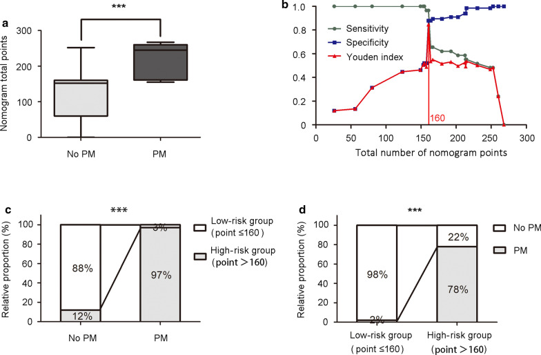
Fig. 5.
Clinical significance of nomogram to predict peritoneal metastasis. a Total number of nomogram points in non-peritoneal metastasis (Mean ± SEM: 118.6 ± 7.93) and peritoneal metastasis (Mean ± SEM: 216.4 ± 8.77) patients were demonstrated by box plot. The box plot shows the full range of variation (error bars: min and max) with the line representing median. b When 160 was set as the cut-off value determined by ROC analysis and Youden index, nomogram had the best sensitivity (0.97) and specificity (0.88). Youden index = Sensitivity + Specificity − 1. c Positive rate (97%), negative rate (88%), false positive rate (12%), and false negative rate (3%) of the nomogram stratified into low-risk group (total number of nomogram points ≤ 160) and high-risk group (total number of nomogram points > 160). d Proportion of patients with or without peritoneal metastasis was demonstrated in low-risk group and high-risk group. PM peritoneal metastasis; ***P < 0.001

