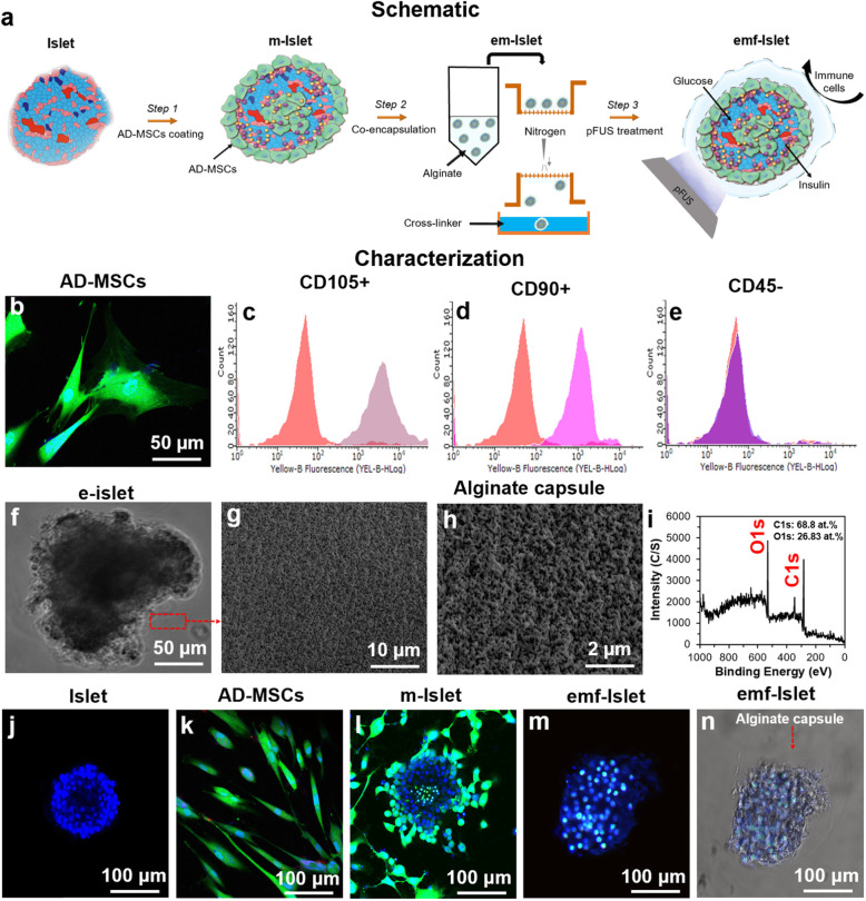
Fig. 1.
Experimental overview and characterization of islets encapsulated with AD-MSCs: a Schematic representation of our three-step approach: step 1: AD-MSC coating, step 2: encapsulation, and step 3: pFUS treatment; characterization of b–e AD-MSCs (b confocal image and c–e FACS analysis), f–i alginate capsule (photographic, SEM images, and XPS scans), and j–n confocal images of an j islet, k AD-MSCs, l islet coated with AD-MSCs, and m–n encapsulated with alginate followed by pFUS treatment. Blue: live cells stained with Hoechst. Green: AD-MSCs stained with FDA

