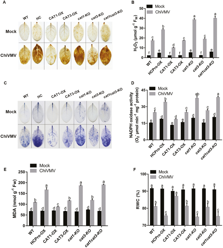
Fig. 6.
Oxidative damage of plants with or without ChiVMV infection. (A) H2O2 levels were detected by DAB staining. (B) Quantitative measurements of H2O2 content. (C) Superoxide contents were detected by NBT staining. (D) Quantitative measurements of NADPH oxidase activity. Quantitative measurements of (E) RWC and (F) MDA content. Systemically infected leaves were collected for detection. Bars represent the mean and SD of values obtained from three biological repeats. Significant differences (P<0.05) are denoted by different lower case letters. FW, fresh weight.

