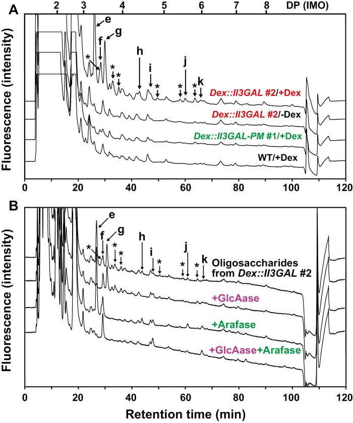
Fig. 5.
Identification of oligosaccharides liberated from type II AGs in vivo. (A) Chromatogram of detected oligosaccharides liberated from type II AGs by the action of Il3GAL. The oligosaccharides in the soluble fraction were derivatized with ABEE and detected by HPLC. Arrows indicate liberated oligosaccharides as follows: e, MeGlcAGal2; f, Gal3; g, AraGal3; h, MeGlcAGal3; i, Gal4; j, MeGlcA-β-1,6-Gal4; k, β-1,6-galactopentaose. Asterisks indicate oligosaccharides released from type II AGs but not assigned. The elution positions of glucose and IMOs with DP 2–8 are indicated on the top. (B) Enzymatic digestion of AG oligosaccharides. The labeled oligosaccharides were digested with GlcAase (+GlcAase), Arafase (+Arafase), or both enzymes (+GlcAase +Arafase). The elution positions of β-1,6-galactooligosaccharides and MeGlcA-β-1,6-galactooligosaccharides are shown in Supplementary Fig. S3.

