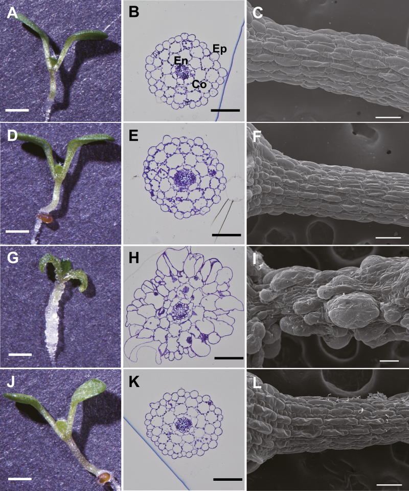
Fig. 6.
Tissue disorganization in the transgenic plants. WT plants treated with Dex (A–C), Dex::Il3GAL plants grown without Dex (D–F), Dex::Il3GAL plants treated with Dex(G–I), and Dex::Il3GAL-PM plants treated with Dex (J–L) were observed. Plants were grown on MS agar medium containing 0.1 μM Dex for 7 d under continuous light before analysis. (A, D, G, J) Stereoscope images of seedlings. Scale bar=1 mm. Plants treated with different Dex concentrations are shown in Supplementary Fig. S4. (B, E, H, K) Cross-sections of the middle region of the hypocotyl stained with toluidine blue O. Scale bar=100 µm. Ep, epidermis; Co, cortex; En, endodermis. (C, F, I, L) SEM images of hypocotyls. Scale bar=100 μm.

