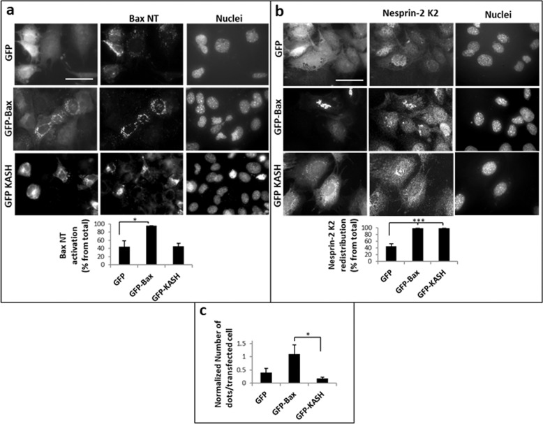
Fig. 7. GFP-KASH-induced nesprin-2 redistribution is insufficient to promote interaction between nesprin-2 and Bax.
Caspase-9−/− MEFs were transfected with GFP, GFP-Bax or GFP-KASH expression vectors. 24 h later, cells were assessed for Bax-NT exposure using anti-Bax (6A7) Ab (a), nesprin-2 localization using anti-nesprin-2 K2 (b) and for interaction between nesprin-2G and Bax using the Duolink-PLA approach (c). The photomicrographs in a and b of each treatment represent the same field visualized separately for detection of GFP fluorescence, Bax-NT or nesprin-2 K2 staining and Hoechst-stained nuclei. Presented results are from a representative experiment (n = 4). Bar = 50 µm. Quantification of the percentage of GFP, GFP-Bax, or GFP-KASH expressing cells, exhibiting Bax-NT exposure or nesprin-2 K2 redistribution from the total population of cells exhibiting GFP fluorescence (total) (at least 50 cells) is shown under the corresponding images. Values are presented as mean ± SEM (error bars) (n = 4). (*p < 0.05, ***p < 0.002; two tailed student’s t test). (c) Quantification of the interaction between nesprin-2G and Bax. Duolink PLA assay was preformed using the Bax (6A7) Ab and anti-nesprin-2G Ab. The results are expressed as the number of dots in transfected cell (at least 20 cells for each treatment) normalized to cell size. Values are presented as mean ± SEM (error bars) (n = 4). (*p < 0.05, two tailed student’s t test between GFP-Bax and GFP-KASH treatments).

